Scientists Expand PET Imaging Options Through Simpler Chemistry
The University of Arizona has developed a new chemical process for radio-labeling PET contrast agents that has now been licensed to startup TheraCea Pharma.

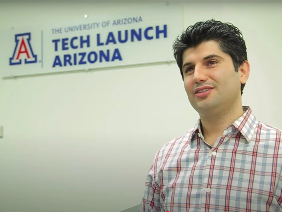
Iman Daryaei, Ph.D.
Tech Launch Arizona/Paul Tumarkin
TUCSON, Ariz. – Researchers at the University of Arizona have developed a rapid, simplified method for producing radio-labeling compounds used for positron emission tomography, also known as PET scans. Iman Daryaei, Ph.D., who led the research and development of the technology, co-invented the method during his doctoral studies in the Department of Chemistry and Biochemistry associated with both the College of Science and the College of Medicine – Tucson.
Working with Tech Launch Arizona, the office of the university that commercializes inventions stemming from research, the team developed the intellectual property and licensed it to startup TheraCea Pharma.
The company plans to package the method as a kit, allowing labs to efficiently do the radio-labeling prior to administering the agents to patients for scans.
“Our advantage is that we’ve simplified the methods that allow non-experts to do the nuclear chemistry,” says Daryaei, TheraCea co-founder and CEO. “We’ve done the work to make sure that it’s useful and simple for any lab technician to use.”
Part of what makes PET imaging work is that specific formulations containing small, safe amounts of a radioactive isotope are injected into the body as contrast agents. But making these agents, which commonly use the radioisotope Flourine-18, is no small task. Before the UArizona team’s innovation, this required an expert in nuclear chemistry to work through the processes to bind Flourine-18 to diagnostic agents and produce a patient-ready formulation.
“Time is of the essence in the preparation and administration of the agents necessary to perform a PET scan,” noted Bruce Burgess, Director, Venture Development for TLA and experienced entrepreneur in the healthcare space. “This is a very real problem that affects the ability of providers to deliver high-quality service. We’re happy to have been able to provide TheraCea with guidance and support throughout the startup process to help the team prepare for a successful launch.”
The new method aims to make PET scans easier and more efficient. To further develop the technology, TheraCea received an SBIR award from the National Institute of Biomedical Imaging and Bioengineering (NIBIB) and the National Institutes of Health (NIH).
“Ultimately, earlier diagnosis means more and better options for treatment,” said Daryaei, “and, ideally, more treatment success.”
A cousin of the CT scan which renders images in two dimensions, PET represents one of the most modern, sensitive imaging techniques available to doctors to image the interior of the human body. With its unmatched detail and sensitivity, combined with the ability to render images in 3D and detect chemical activity, PET offers the ability to detect, map and monitor cancer better and at an earlier stage of disease than ever before.
Hospitals across the U.S. house about 2,500 PET scanners and perform about 2 million scans annually.

