
Tech Blog: UA’s Kostuk and Barton Develop Method and Device to Improve Screening for Ovarian Cancer
Ovarian cancer only accounts for about three percent of cancers among women, but results in the highest number of deaths when compared to all other cancers involving the female reproductive system [1]. The American Cancer Society estimates that nearly 22,280 women will receive a new ovarian cancer diagnosis in 2016. Of those women, approximately 14,240 will die from the disease [2]. Unfortunately, current screening and diagnostic abilities result in seventy percent of affected women being diagnosed with stage III or IV cancer, a widespread intra-abdominal disease. Because the dismal outlook for these women so highly relies on screening and diagnostic abilities, the introduction of new technology capable of reliably diagnosing early stage ovarian cancer could help reduce the morbidity, mortality, and large economic impact of this disease [3].
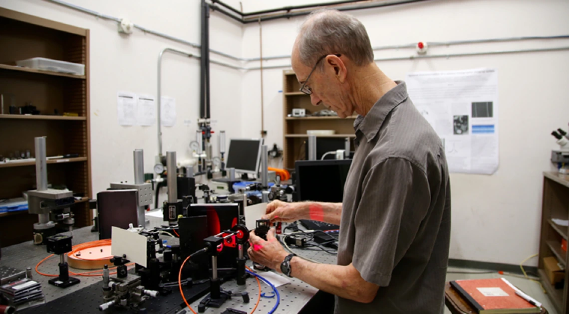
Raymond K. Kostuk Ph.D., of the Department of Electrical and Computer Engineering in the College of Engineering and the College of Optical Sciences, and Jennifer Barton Ph.D., of the Department of Biomedical Engineering and Interim Director of the BIO5 Institute(link is external), have dedicated their recent research to a novel method and device for medical imaging, specifically imaging for detection and diagnosis of ovarian cancer, based on a volume holographic imaging system (VHIS). Kostuk and Barton’s VHIS is built to “simultaneously view micro-meter size cell and tissue features at and below the tissue surface.”[3] This imaging system is modular by design, has interchangeable components, and offers two illumination options.
“The VHIS approach does not require scanning and forms multiple images at camera frame rates. In addition, because the hologram also disperses light, the VHIS method also obtains spectral information about the sample providing a means for fluorescence imaging with biomarkers. All of this can lead to sophisticated, very low-cost (1/5th -1/10th the cost of similar instruments), medical instruments for identifying cancerous tissue at an early age,” said Kostuk [3].
Kostuk and Barton have successfully completed a clinical trial using a “bench-top” version of the system to view all depths of a surgically removed ovarian tissue sample and are currently completing development of a handheld rigid endoscope version of the imaging system. When describing his work on the handheld version, Kostuk said it has “passed the sterilization and clinical engineering certifications and has been used in two surgeries, which validates the ability to reduce this system to a handheld device capable of laparoscopic surgical procedures.” [3]
Select the links below to learn more about these available technologies:
- Confocal Rainbow Volume Holographic Imaging System (UA11-034)
- Dual Hologram Confocal Rainbow Volume Holographic Imaging System (UA11-076)
- Volume Holographic Imaging System VHIS Endoscope (UA12-016)
- Optical Design for the Volume Holographic Imaging System VHIS Endoscope (UA12-072)
Sources:
[1] Ovarian Cancer. (n.d.). Retrieved August 15, 2016, from http://www.cancer.org/cancer/ovariancancer/index(link is external)
[2] What are the key statistics about ovarian cancer? (2016, February 4). Retrieved August 15, 2016, from http://www.cancer.org/cancer/ovariancancer/detailedguide/ovarian-cancer-key-statistics(link is external)
[3] Kostuk, R. (2016, August 11). Docx.
Photo: Raymond K. Kostuk, Ph.D., exhibits the bench-top VHIS at work in his lab located in the University of Arizona’s Department of Electrical and Computer Engineering. Credit: Taylor Hudson/Tech Launch Arizona